Home

Pygmalion wheat cheek gfp labeled cells unused Association Lake Taupo

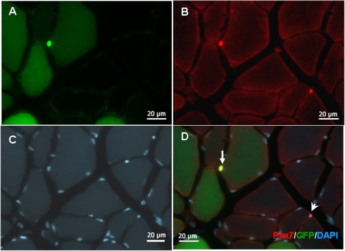
Options for tracking GFP-Labeled transplanted myoblasts using in  vivofluorescence imaging: implications for tracking stem cell fate | BMC  Biotechnology | Full Text
Options for tracking GFP-Labeled transplanted myoblasts using in vivofluorescence imaging: implications for tracking stem cell fate | BMC Biotechnology | Full Text

Prolonged Dormancy and Site-specific Growth Potential of Cancer Cells  Spontaneously Disseminated from Nonmetastatic Breast Tumors as Revealed by  Labeling with Green Fluorescent Protein | Clinical Cancer Research
Prolonged Dormancy and Site-specific Growth Potential of Cancer Cells Spontaneously Disseminated from Nonmetastatic Breast Tumors as Revealed by Labeling with Green Fluorescent Protein | Clinical Cancer Research

Category: Microscopy. Image description: Human keratinocyte cells expressin GFP  labeled keratin-14 (green) stained for DNA (blue). Therape… | Biology, Bio  art, Cell
Category: Microscopy. Image description: Human keratinocyte cells expressin GFP labeled keratin-14 (green) stained for DNA (blue). Therape… | Biology, Bio art, Cell

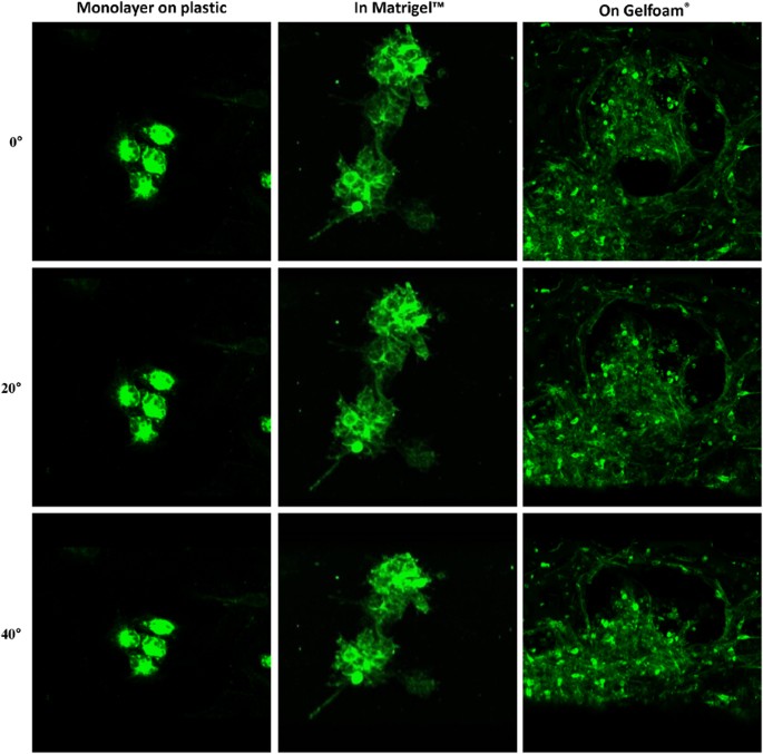
Fluorescence Tracked Green Fluorescent Protein (GFP)-Labeled... | Download  Scientific Diagram
Fluorescence Tracked Green Fluorescent Protein (GFP)-Labeled... | Download Scientific Diagram

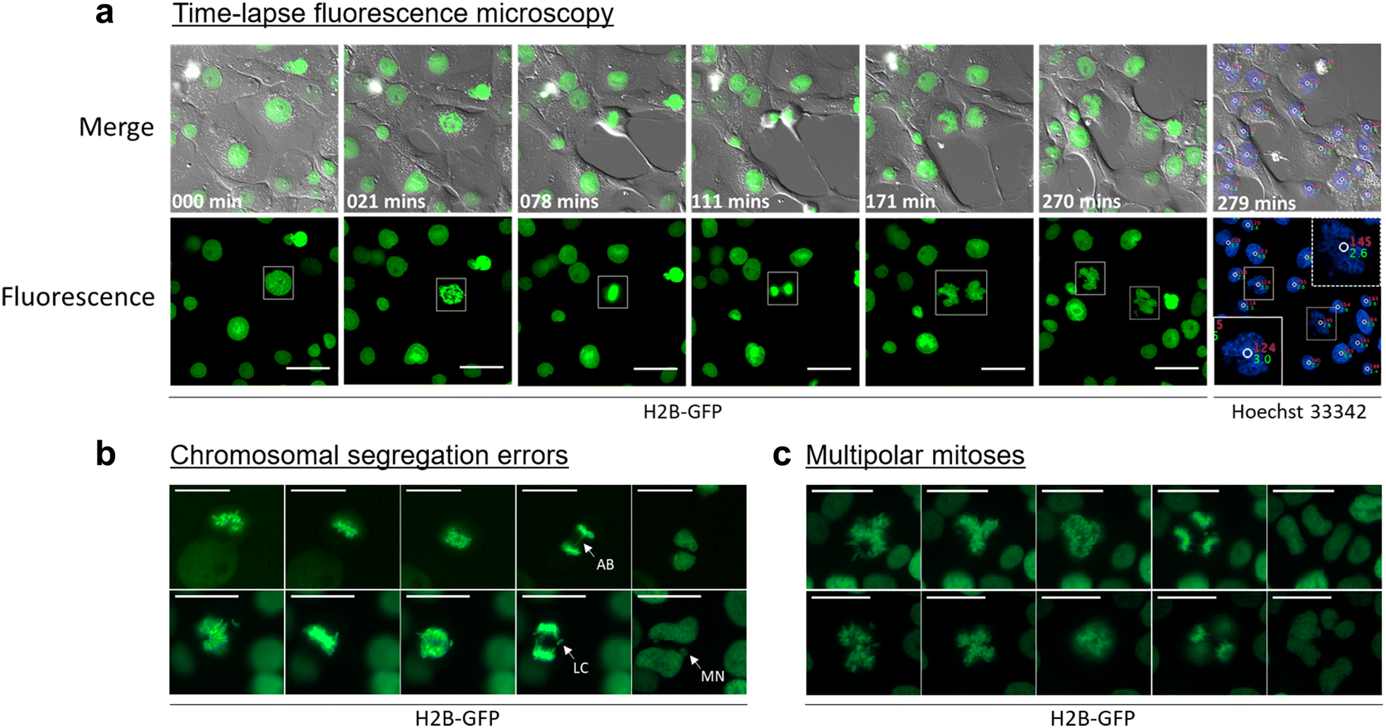
GFP labeling allows in vitro cell tracking. (a) Live fluorescence... |  Download Scientific Diagram
GFP labeling allows in vitro cell tracking. (a) Live fluorescence... | Download Scientific Diagram

Labeling Neural Cells Using Adenoviral Gene Transfer of Membrane-Targeted  GFP: Neuron
Labeling Neural Cells Using Adenoviral Gene Transfer of Membrane-Targeted GFP: Neuron

Cellular Dynamics Visualized in Live Cells in Vitro and in Vivo by  Differential Dual-Color Nuclear-Cytoplasmic Fluorescent-Protein Expression  | Cancer Research
Cellular Dynamics Visualized in Live Cells in Vitro and in Vivo by Differential Dual-Color Nuclear-Cytoplasmic Fluorescent-Protein Expression | Cancer Research

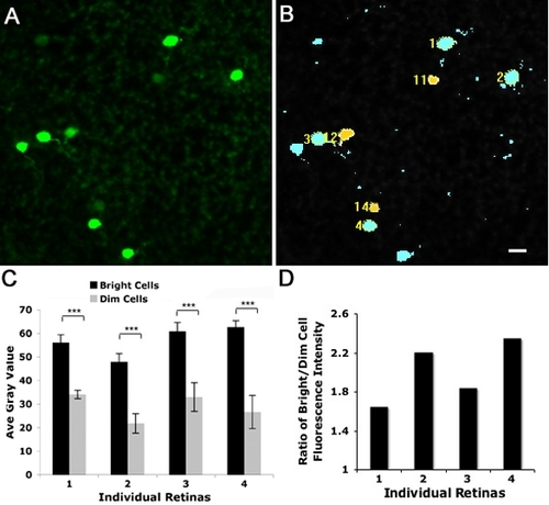
PLOS ONE: Fluorescent Labeling of Newborn Dentate Granule Cells in GAD67-GFP  Transgenic Mice: A Genetic Tool for the Study of Adult Neurogenesis
PLOS ONE: Fluorescent Labeling of Newborn Dentate Granule Cells in GAD67-GFP Transgenic Mice: A Genetic Tool for the Study of Adult Neurogenesis

Application of GFP imaging in cancer | Laboratory Investigation

Figure 1 from Tracking of green fluorescent protein (GFP)-labeled LAK cells  in mice carrying B16 melanoma metastases. | Semantic Scholar
Figure 1 from Tracking of green fluorescent protein (GFP)-labeled LAK cells in mice carrying B16 melanoma metastases. | Semantic Scholar
EGFP-labeled cells co-express myeloid lineage markers. Dual... | Download  Scientific Diagram
EGFP-labeled cells co-express myeloid lineage markers. Dual... | Download Scientific Diagram

In vivo tracking of T cell development, ablation, and engraftment in  transgenic zebrafish | PNAS
In vivo tracking of T cell development, ablation, and engraftment in transgenic zebrafish | PNAS

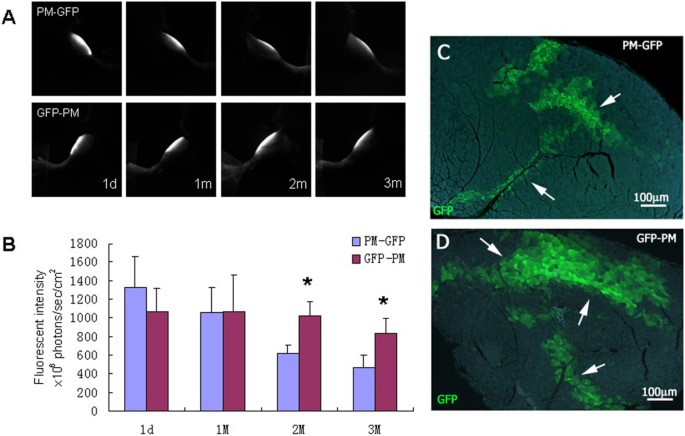
Options for tracking GFP-Labeled transplanted myoblasts using in  vivofluorescence imaging: implications for tracking stem cell fate | BMC  Biotechnology | Full Text
Options for tracking GFP-Labeled transplanted myoblasts using in vivofluorescence imaging: implications for tracking stem cell fate | BMC Biotechnology | Full Text
GFP labeling for engrafted cell following after injection into the... |  Download Scientific Diagram
GFP labeling for engrafted cell following after injection into the... | Download Scientific Diagram

JCI - Identifying local and descending inputs for primary sensory neurons

ZFIN Figure: Meng et al., 2008, Fig. 5

GFP for in vivo imaging of subcellular structures in plant cells: Trends in  Plant Science
GFP for in vivo imaging of subcellular structures in plant cells: Trends in Plant Science

Figure 18, [Profiling GFP-labeled embryonic cells. Intact...]. - WormBook -  NCBI Bookshelf
Figure 18, [Profiling GFP-labeled embryonic cells. Intact...]. - WormBook - NCBI Bookshelf

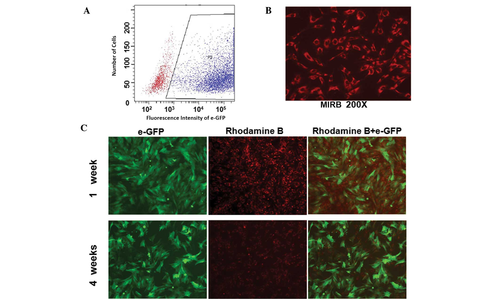
Assessment of biological characteristics of adipose tissue‑derived stem  cells co‑labeled with Molday ION Rhodamine B™ and green fluorescent protein  in vitro
Assessment of biological characteristics of adipose tissue‑derived stem cells co‑labeled with Molday ION Rhodamine B™ and green fluorescent protein in vitro

Prostate progenitor cells proliferate in response to castration -  ScienceDirect
Prostate progenitor cells proliferate in response to castration - ScienceDirect

Cell-type–specific, multicolor labeling of endogenous proteins with split  fluorescent protein tags in Drosophila | PNAS
Cell-type–specific, multicolor labeling of endogenous proteins with split fluorescent protein tags in Drosophila | PNAS

Measuring DNA content in live cells by fluorescence microscopy | Cell  Division | Full Text
Measuring DNA content in live cells by fluorescence microscopy | Cell Division | Full Text

Confocal images of Spider-GFP labeled cells on the vent | Open-i

Diffusible GRAPHIC to visualize morphology of cells after specific cell–cell  contact | Scientific Reports
Diffusible GRAPHIC to visualize morphology of cells after specific cell–cell contact | Scientific Reports